Diffusion MRI
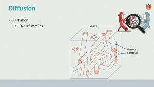
This lecture provides a comprehensive introduction to Diffusion MRI, a quantitative imaging technique that measures the microscopic random motion of water molecules (Brownian motion) within brain tissue. The lecture explains how MRI uses diffusion-sensitizing gradients to create signal loss in mobile water molecules, allowing researchers to calculate the Apparent Diffusion Coefficient (ADC) and visualize tissue microstructures. The lecture covers the progression from basic diffusion-weighted imaging (DWI) to advanced Diffusion Tensor Imaging (DTI), detailing how these methods are used to assess brain development (myelination), detect pathologies like strokes (cytotoxic edema) and tumors, and map white matter pathways through tractography.
Learning objectives:
By the end of this lecture, students will be able to:
- Explain how water molecule movement is influenced by temperature, viscosity, and physical barriers like cell membranes
- Describe how moving molecules in a magnetic gradient leads to signal reduction, forming the basis of diffusion contrast
- Understand the relationship between B-values, signal intensity, and the calculation of ADC maps
- Differentiate between isotropic and anisotropic diffusion, and understand parameters like Fractional Anisotropy (FA)
- Physics of Diffusion:
- Introduction to T2-weighted reference images (B0) and the fast Echo Planar Imaging (EPI) technique
- How myelination changes T1, T2, and ADC values in infants compared to adults
- Diffusion Tensor Imaging (DTI): * Measuring diffusion in 3D space using at least six gradient directions
- The concept of the diffusion "ellipsoid" (cigar-shaped vs. pumpkin-shaped)
- Tractography: * Deterministic vs. Probabilistic: Methods for following fiber directions to map the brain's "highways" like the corpus callosum and corticospinal tract
- Artifacts: Addressing geometric distortions and susceptibility artifacts inherent in fast-diffusion scans.