MS Case-based diagnostics
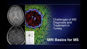
This lecture features a clinical lecture focused on the practical application of the McDonald criteria for diagnosing Multiple Sclerosis (MS) through real-world patient cases. The lecture reviews key imaging landmarks, such as periventricular, juxtacortical, and infratentorial lesions, while emphasizing the importance of dissemination in space (DIS) and dissemination in time (DIT). Through several detailed case studies, the session explores the diagnostic journey from initial presentation to long-term management, including the nuances of Radiologically Isolated Syndrome (RIS), the impact of optic neuritis, and the critical decision-making process behind selecting first-line versus high-efficacy therapies.
Learning objectives
By the end of this lecture, students will be able to:
- Identify the typical anatomical localizations for MS lesions on MRI
- Understand the core diagnostic requirements of dissemination in space and time under current and evolving guidelines
- Differentiate MS from its "cousins" and differential diagnoses like ADEM, NMOSD, and vascular disease through lesion morphology and clinical presentation
- Evaluate the risks and benefits of various MS treatment tiers, from oral medications to high-efficacy infusions and stem cell therapy.
- Recognize the clinical significance of Radiologically Isolated Syndrome (RIS) and its progression to symptomatic MS.
- Monitor treatment safety, specifically tracking white blood cell counts to prevent infections like PML during high-efficacy therapy.
- Diagnostic Framework
- Advanced Imaging Markers: The role of Optical Coherence Tomography (OCT) in measuring retinal nerve fiber layer thinning and the Central Vein Sign on MRI
- Case Studies
- Symptoms Management: A brief discussion on the complex origins of MS fatigue and its lack of a direct radiological correlate